Home
/ Blood Cells Under Microscope, Red Blood Cell Analysis Using Ultraviolet Microspectroscopy : Primary job is to carry oxygen.
Blood Cells Under Microscope, Red Blood Cell Analysis Using Ultraviolet Microspectroscopy : Primary job is to carry oxygen.
Blood Cells Under Microscope, Red Blood Cell Analysis Using Ultraviolet Microspectroscopy : Primary job is to carry oxygen.. Scanning electron microscopes generate similar images. Red blood cells also have a nucleus during their stages of development. This beautiful watercolor painting depicts blood smears of different blood cells ready to be analyzed. Red blood cells (rbcs) as seen under the microscope in isotonic, hypotonic and hypertonic solutions. · view the slide under the microscope (starting with 10x magnification).
Nrbcs are the precursor to red blood cells and they come from the bone marrow. Find the perfect blood cells microscope stock photos and editorial news pictures from getty images. After you get the mrna vaccine, the membrane of red blood cells becomes abnormal and they clump together. Blood cells under the microscope! With the high power roachscope we can get a closer look at the cells that compose us.
Red Blood Cells Under Microscope Poster By Avidfan2000 Redbubble from ih1.redbubble.net 6 blood cells watercolor print histology and hematology art | etsy. A few white blood cells can also be seen with the red. Red blood cells (rbcs) as seen under the microscope in isotonic, hypotonic and hypertonic solutions. Unlike other blood cells (which may leave the vessels to carry out their functions), red blood cells remain within the vascular network from where they are transported throughout the body. The revelation was pretty shocking. · view the slide under the microscope (starting with 10x magnification). Find the perfect blood cells microscope stock photos and editorial news pictures from getty images. In case of eneven distribution of blood cells.
Since this occurs in the bone marrow we do not normally see these nucleated red.
A few white blood cells can also be seen with the red. Since this occurs in the bone marrow we do not normally see these nucleated red. 6 blood cells watercolor print histology and hematology art | etsy. It is why human red blood cells, under the microscope, do not show any nucleus. Higher magnifications show the individual cells and it is even possible to observe the fact that red blood corpuscles do not have a nucleus. All cells need a nucleus for replication and maturation. Plasma constitutes about 55% of. Obtained by placing the slides under a compound or optical microscope under illumination blood cell. Under microscopic examination, red blood cells are a biconcave disk in shape and have a flattened center. The red blood cell is the most abundant blood cell in the body (about 45%). Different component of blood cells like eosonophils, basophiles and neutrophils have different morphological demarcations and are therefore very easy to distinguish under a microscope. Human blood contains many different components, from white blood cells to platelets, but the most abundant component by far are red blood cells. Some people saw the images of red blood cells under the microscope.
Blood cells under the microscope. Plasma constitutes about 55% of. It is graduated to dilute blood 1 in 100 or 1 in 200. With the high power roachscope we can get a closer look at the cells that compose us. Images related to blood cancer.
Light Microscope Images Of Giemsa Stained Red Blood Cell Samples A Download Scientific Diagram from www.researchgate.net 4k00:14blood under a microscope, the movement of red blood cells, cells that carry oxygen throughout the human body 4k. Different component of blood cells like eosonophils, basophiles and neutrophils have different morphological demarcations and are therefore very easy to distinguish under a microscope. • contents of blood blood contains three main components and several sub components that do everything from carry oxygen throughout the body to clot when there. Blood analysis advertising poster realistic design with test tube filled blood and cell in red human liquid under multiple zooming. All cells need a nucleus for replication and maturation. Your doctor may also perform a blood smear, which is a way of looking at your blood cells under the microscope. Red blood cells also have a nucleus during their stages of development. Unlike other blood cells (which may leave the vessels to carry out their functions), red blood cells remain within the vascular network from where they are transported throughout the body.
Blood cells under the microscope!
Blood cells show clear demarcations under the microscope. What to keep in mind? Blood cells under the microscope. Blood cells under a microscope. More info on cell staining. Blood cells types infographics with red pipe arteria illustration. Red blood cells (rbcs) as seen under the microscope in isotonic, hypotonic and hypertonic solutions. Under microscopic examination, red blood cells are a biconcave disk in shape and have a flattened center. The red blood cell is the most abundant blood cell in the body (about 45%). In case of eneven distribution of blood cells. Unlike other blood cells (which may leave the vessels to carry out their functions), red blood cells remain within the vascular network from where they are transported throughout the body. Pdf | white blood cells (wbc) play a significant role in the immune system by protecting the body these images are. A few white blood cells can also be seen with the red.
Movement of red blood cells in an artery under the microscope. You will use a lancet to safely draw blood from your fingertip in order to view blood cells under the roachscope. 6 blood cells watercolor print histology and hematology art | etsy. Red blood cells also have a nucleus during their stages of development. More info on cell staining.
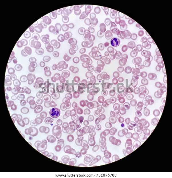
Human Blood Smear Under 100x Light Stock Photo Edit Now 751876783 from image.shutterstock.com Blood cells microscope stock images from offset. Your doctor may also perform a blood smear, which is a way of looking at your blood cells under the microscope. Scanning electron microscopes generate similar images. Your blood is a soup of different kinds of cells, and around 1 % of these cells are immune cells also known as white blood cells. Hi, here is an image of some blood cells under the microscope. The revelation was pretty shocking. Nrbcs are the precursor to red blood cells and they come from the bone marrow. They usually hit the bloodstream in anemia cases, thalassemia in which, just to let you know there is an nrbc in the center between the 4 and 5 in the microscope.
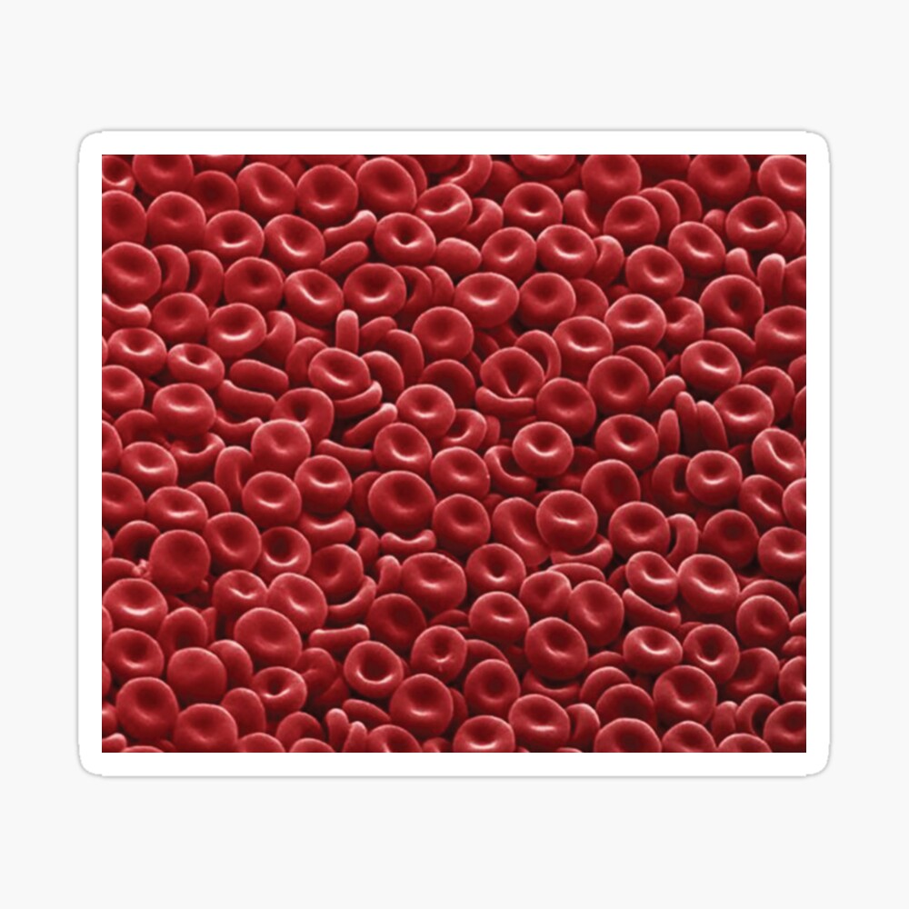
I think it is seamless so should be able to tiled into a much larger picture.
Under microscopic examination, red blood cells are a biconcave disk in shape and have a flattened center. The red blood cell is the most abundant blood cell in the body (about 45%). Different component of blood cells like eosonophils, basophiles and neutrophils have different morphological demarcations and are therefore very easy to distinguish under a microscope. And the bottom, this white blood cell is a segmented. Blood cells microscope stock images from offset. It is graduated to dilute blood 1 in 100 or 1 in 200. After you get the mrna vaccine, the membrane of red blood cells becomes abnormal and they clump together. Killer t cells are immune cells that can kill certain other cells, including foreign cells, cancer. This is what the human body really looks like under a microscope. Images related to blood cancer. Your doctor may also perform a blood smear, which is a way of looking at your blood cells under the microscope. Live blood cell analysis a short description of live blood cell analysis under a microscope. 6 blood cells watercolor print histology and hematology art | etsy.
